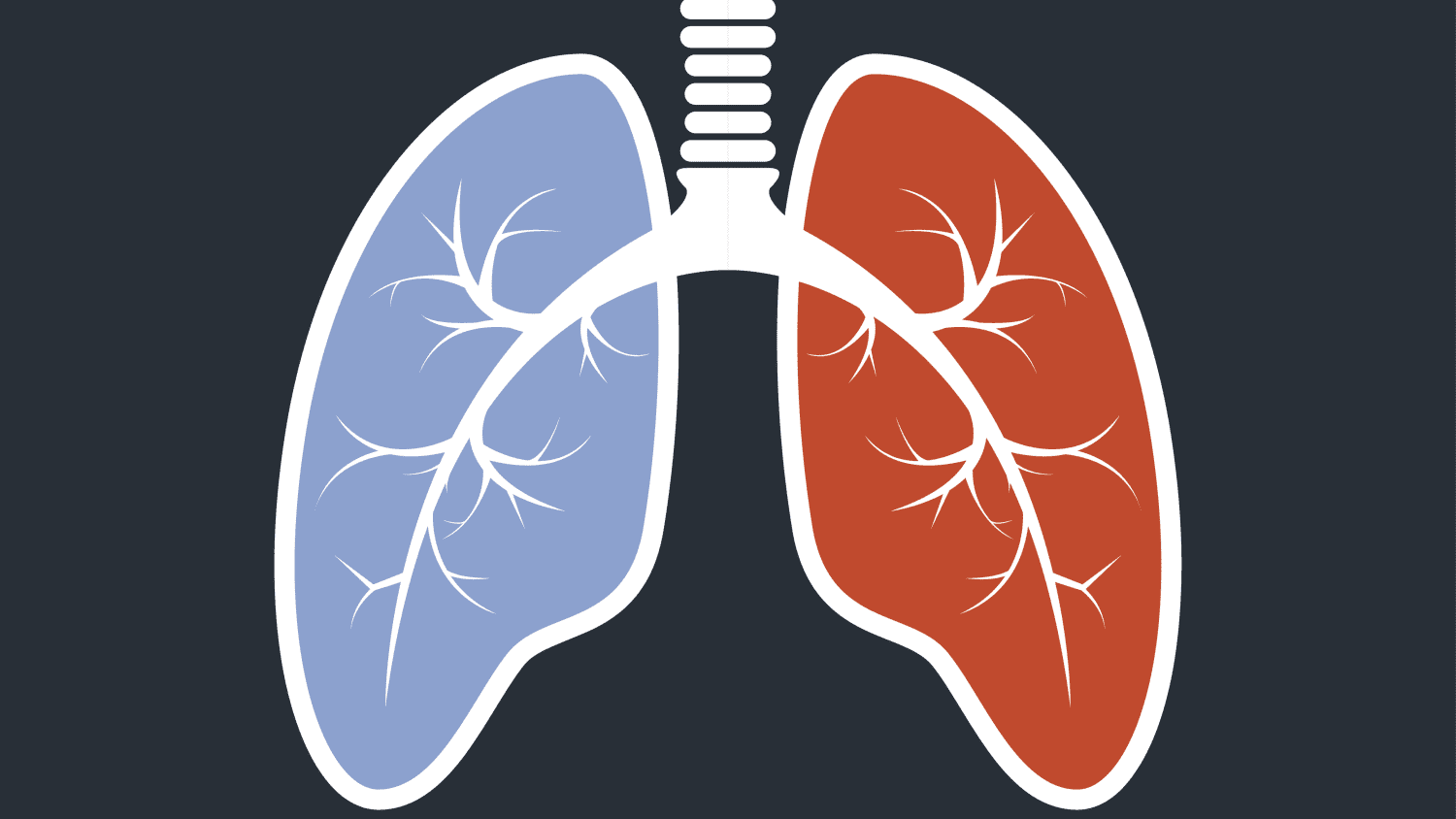
What is Pulmonary Vein Stenosis? Medical science has not pinpointed the exact cause of pulmonary vein stenosis yet. But experts think this condition may be related to a heart valve problem or issues with blood flow. Surgery is sometimes necessary for people suffering from pulmonary vein stenosis to treat it. Please look at these top facts on pulmonary vein stenosis and its treatment.
Pulmonary vein stenosis (PVS) blocks one or both prominent veins that carry blood from your heart to your lungs. This is a life-threatening condition, as the narrowed area prevents the heart’s blood from reaching your lungs. The resulting shortness of breath can be caused by exercise, heavy breathing, or even a sudden burst of activity.

What is pulmonary vein stenosis?
Pulmonary vein stenosis is characterized by an abnormally narrow opening in the heart. Pulmonary vein stenosis is a condition with a tiny space in the left atrium. This can result from narrowing the pulmonary veins or the gap between the left atrium and left ventricle.
The right heart receives blood from the body via the superior vena cava (SVC). From the right atrium, the blood passes through a valve called the mitral valve into the left atrium. From there, the blood flows into the left ventricle, where it is pumped out to the rest of the body.
What are the symptoms of pulmonary vein stenosis?
Pulmonary vein stenosis is narrowing pulmonary veins due to progressive scarring. Symptoms of this condition may include chest pain, shortness of breath, and a blood clot in the lungs. People may also feel dizzy when standing up and experience low oxygen levels when resting. The symptoms of pulmonary vein stenosis can vary from person to person. Not everyone who develops pulmonary vein stenosis will have the same symptoms.
Things you should keep in your Mind
- What are the symptoms of pulmonary vein stenosis?
- What causes pulmonary vein stenosis?
- How is pulmonary vein stenosis diagnosed?
- What are the treatment options for pulmonary vein stenosis?
- What are the complications of pulmonary vein stenosis?
- How common is pulmonary vein stenosis?
- What are the long-term effects of pulmonary vein stenosis?
How is pulmonary vein stenosis diagnosed?
A diagnosis of pulmonary vein stenosis is typically made after an echocardiogram or cardiac catheterization. The first step is an echocardiogram to diagnose pulmonary vein stenosis to determine abnormal heart valves. Your echocardiogram will look at the size and shape of your heart and its valves, as well as the strength of your heart muscle. This test may help determine the severity of your disease and plan a treatment that is most likely to benefit you.
It will also evaluate how well your heart’s left and right sides work together during each heartbeat. Your doctor will place small devices, or catheters, in the arteries of your legs. A thin tube attached to the catheter will be threaded into your heart and hooked up to a device that measures blood flow through your heart. Heart rhythm problems are common in people with HIV. This includes atrial fibrillation (AFib), in which the upper chambers of your heartbeat are too fast, and the lower sections are too slow.
What are the treatment options for pulmonary vein stenosis?
Pulmonary vein stenosis occurs when the pulmonary veins are narrowed or blocked. The treatment options for pulmonary vein stenosis vary based on the severity of the condition. Patients with less severe cases may not require any treatment, while more severe cases require surgery. If surgery is needed, the goal is to relieve the obstruction to blood flow, which will allow the patient to have adequate breathing. There are several different surgical techniques for treating pulmonary vein stenosis. One type of surgery is called the clipart or a pulmonary venous patch.
Is pulmonary vein stenosis preventable?
Pulmonary vein stenosis is an ailment that affects the heart’s left ventricle. It is caused by inflammation of the pulmonary vein valves and is often found in high blood pressure adults. Pulmonary vein stenosis is a heart problem often associated with high blood pressure. If you have pulmonary vein stenosis, your heart needs to work harder to pump blood through your body.
The extra effort can lead to fatigue and shortness of breath. If you have heart failure, your heart isn’t able to pump as well as it used to. Heart failure occurs when the heart is enlarged or damaged. Your heart might not be able to fill with blood, which limits its ability to pump blood through your body. Pulmonary embolism blocks an artery in the lung (the pulmonary artery). This can happen if a blood clot gets dislodged from inside your body and travels through your blood vessels to your lung.
Causes of Pulmonary Vein Stenosis
Veins of the right atrium are prone to stenosis. This condition is caused by narrowing the vein due to disease or injury. The causes of pulmonary vein stenosis are mainly unknown. One theory is that inflammation in the right atrium causes thickening of the vein walls, causing them to stick together.
The other theory is that the vein walls stick together because of scarring and stiffening caused by the inflammation. When inflammation becomes chronic, it causes damage to the heart’s tissue-making it more likely that the same problem will happen again. The right atrium collects before being pumped out of the soul into the arteries. The left atrium is where the blood contains while the heart is pumping.
The prognosis for Pulmonary Vein Stenosis
Pulmonary vein stenosis is a severe heart condition that causes too much blood to flow through the lungs. A person with pulmonary vein stenosis will have difficulty breathing and often has a rapid heart rate.
They may also experience shortness of breath, chest pain, or tightness in their chest. They may not be able to talk or breathe as easily. If a person has severe pulmonary vein stenosis, they may need to be hospitalized. They will be connected to a machine that helps them breathe during their hospital stay.
Prevention Strategies for Pulmonary Vein
The pulmonary vein is the vein in the lungs that drains deoxygenated blood from the alveoli to the heart. The prevention strategies for pulmonary veins are pharmaceuticals to prevent blood clots in the veins. Another method is to prevent clots from breaking loose and traveling through the blood vessels. Several drugs can be used to treat venous thromboembolism.
There are also non-pharmaceutical interventions that can be used to prevent blood clots. These pharmaceuticals are coumarin derivatives, heparin, and Factor VIII. For example, a person should limit their time on the leg press since this exercise increases the pressure in the veins and may cause venous thromboembolism. Some studies have found that wearing compression stockings (see below) can be beneficial for the prevention of DVT and PE.
Risk factors for pulmonary vein stenosis
Pulmonary vein stenosis is a condition where the pulmonary veins are narrowed. This occurs due to arteriosclerosis of the left side of the heart, which restricts blood flow to the lungs. As a result, patients with left-sided heart failure may have shortness of breath and fatigue that worsens with exertion.
Impaired gas exchange leads to fatigue, shortness of breath, and inadequate energy during exertion. As the disease progresses, pulmonary hypertension may develop, which increases the work of the heart relative to the smaller amount of blood it can pump. This can lead to heart failure with less than 40% ejection fraction.
What can you do if you think you have pulmonary vein stenosis?
The right side of the heart has to pump blood through narrow pulmonary veins to the lungs. The left side of the heart pumps blood through large vessels to the rest of the body. The right side of the heart is used more than the left side. When a person breathes out, the right side of the heart contracts to pump blood to the lungs.
During exercise, the right side of the heart pumps harder to deliver more oxygen to the muscles. The right side also plays a more significant role in digestion and elimination. The right side of the heart is about 20% larger than the left. As a result, it requires more energy to pump blood through the body.
Conclusion
Doctors make a small incision in the artery near the ankle and insert a small tube called a catheter into the street. The catheter is then passed into the heart through the route and up to the pulmonary vein, from which blood can be withdrawn or injected to change the pressure in the heart. Sometimes the pressure in the heart becomes too high. This condition is called hypertrophic cardiomyopathy. A doctor can inject the heart with a substance that causes it to contract to reduce stress.


